
Product
ZEISS Intraoperative Fluorescence Technologies
Discovering the unseen
Fluorescence is the property of atoms and molecules called fluorophores to absorb light of a specific wavelength and then emit light of a longer wavelength. Fluorescence microscopy can be based on autofluorescence or the addition of fluorescent dyes. 1,2 Fluorescent dyes can be invisible under normal light. However, a surgical microscope with integrated fluorescence technology illuminates the dye to visualize tumor tissue or blood vessels during surgery.
- Tumor
- Vein
- Reconstructor
Deeper insights. Greater control.
The first use of intraoperative fluorescence imaging during surgery dates back to 1948, when surgeons used intravenous fluorescein to enhance intracranial neoplasms during neurosurgery. 2,9 Since then, additional fluorescent agents have been used for a variety of surgical applications. 4,5,6,9 Intraoperative fluorescence imaging offers the advantages of high contrast and sensitivity, absence of ionizing radiation, ease of use, safety, and high specificity. 7,8,9 Compared to standard unaided vision using white light imaging, real-time fluorescence imaging is helpful in visualizing cancerous tissue and delineating tumor margins. 8 What's more, better visualization of cancer can reduce damage to important normal structures such as nerves, blood vessels, ureters and bile ducts. 9
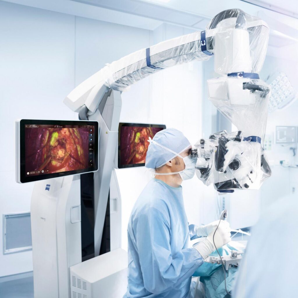
In demanding microsurgery, surgical imaging aids are essential for making the right decisions at the right time. Intraoperative Fluorescence Technologies from ZEISS 1 of gives you the tools you need.
Intraoperative visualization of fluorescently stained structures in tumor surgery
With ZEISS BLUE 400 and ZEISS YELLOW 560
Intraoperative imaging of cerebral blood flow in vascular surgery
With ZEISS INFRARED 800 and ZEISS FLOW 800
Intraoperative fluorescence imaging in reconstructive surgery
With ZEISS INFRARED 800 and ZEISS FLOW 800
Contact us!
Learn more about the product and availability in your country!